Image of migrating cells forming the lateral line in zebrafish. Image courtesy of Julia Peloggia/Piotrowski Lab.
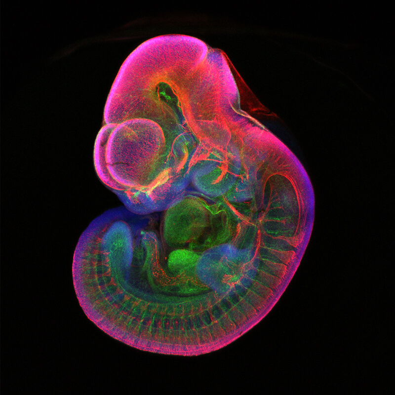
A mouse embryo with fluorescently labeled nerve cells (red) and blood vessels (green). Image courtesy of Melesio Cristobal-Luna/Trainor Lab.
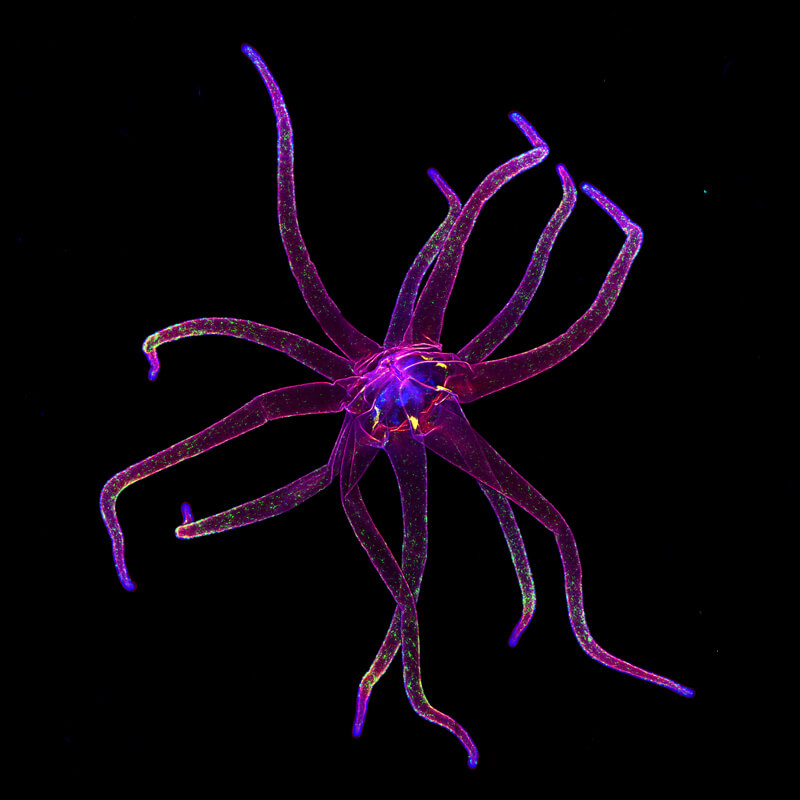
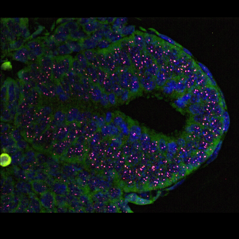
The head region of a sea anemone, with neurons labeled with fluorescent protein. Despite its seemingly simple body plan, the sea anemone possesses a rather sophisticated cellular composition. Image courtesy of Shuonan He/Gibson Lab.
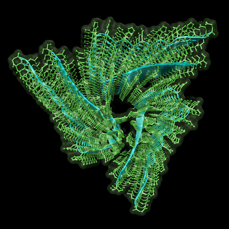
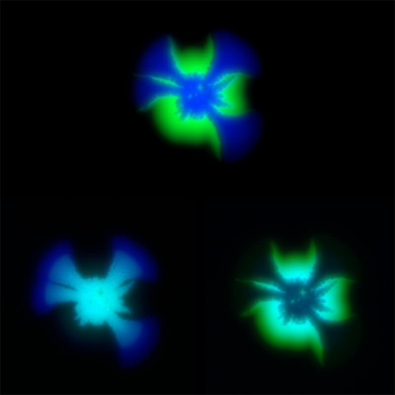
Three dimensional atomic organization of the neuronal protein involved in memory persistance. Image courtesy of Ruben Hervas Millan, Ph.D./Si Lab.
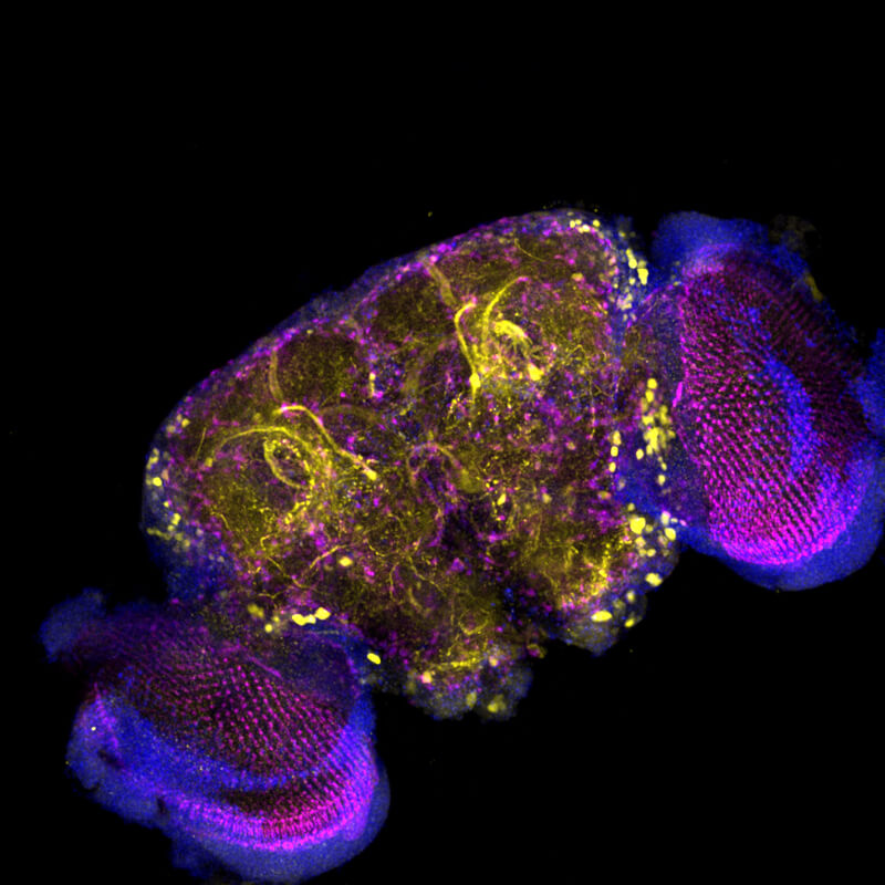
In this adult fruit fly brain imaged on a confocal microscope, sweet-sensing neurons are seen in yellow, while magenta shows the neuronal connections. Image courtesy of Consuelo Perez Sanchez/Si Lab.
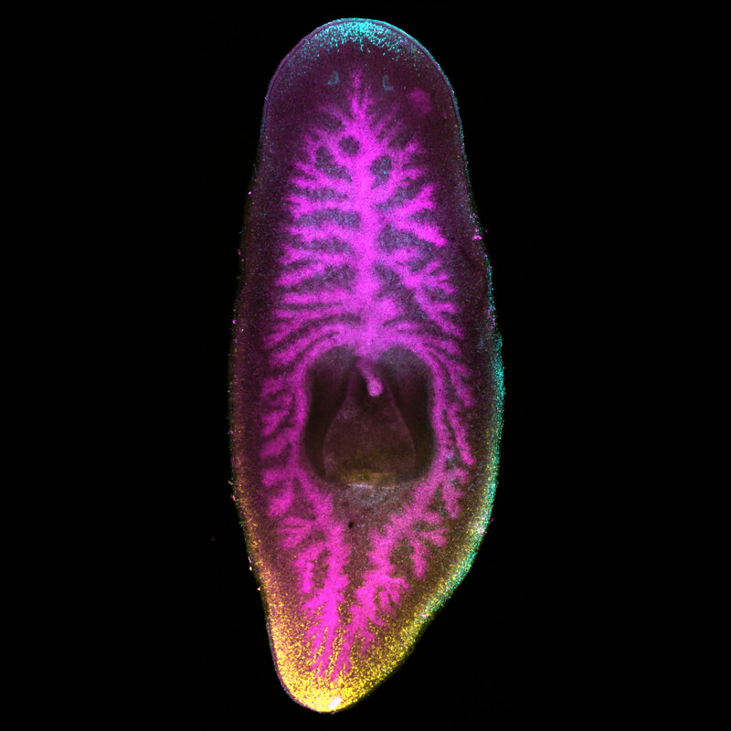
Three color FISH image of the planarian flatworm. porcupine in magenta marks the gut branches. frizzled in yellow is a Wnt receptor which localizes specifically to the posterior of the animal. sfrp1 in cyan is a Wnt inhibitor which is restricted to the anterior of the animal. Image courtesy of Viraj Doddihal/Sanchez Alvarado Lab.
Neuron fibers in a mouse olfactory bulb, a part of the brain that receives sensations of smell. The various colors depict different nerve fibers and help visualize the organization of the mouse nervous system. Image courtesy of Jae Hyoun Seiler/Yu Lab.
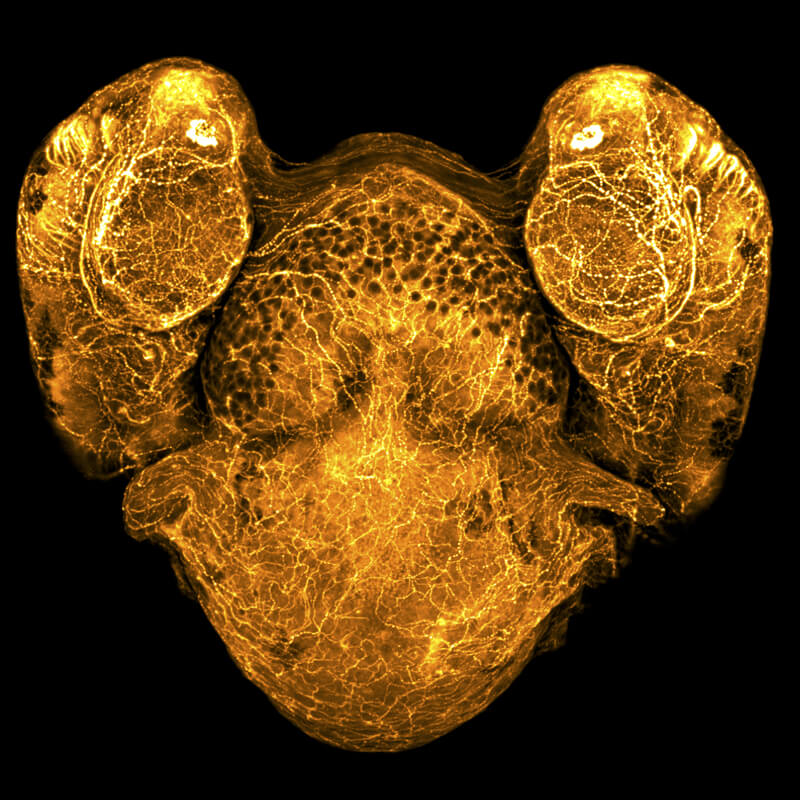
Zebrafish twins. Rare case of spontaneous axis duplication of a two days post fertilization zebrafish embryo. Two heads and bodies share the same heart and intricate axonal connections (orange). Image courtesy of Julia Peloggia de Castro/Sanchez Alvarado Lab.
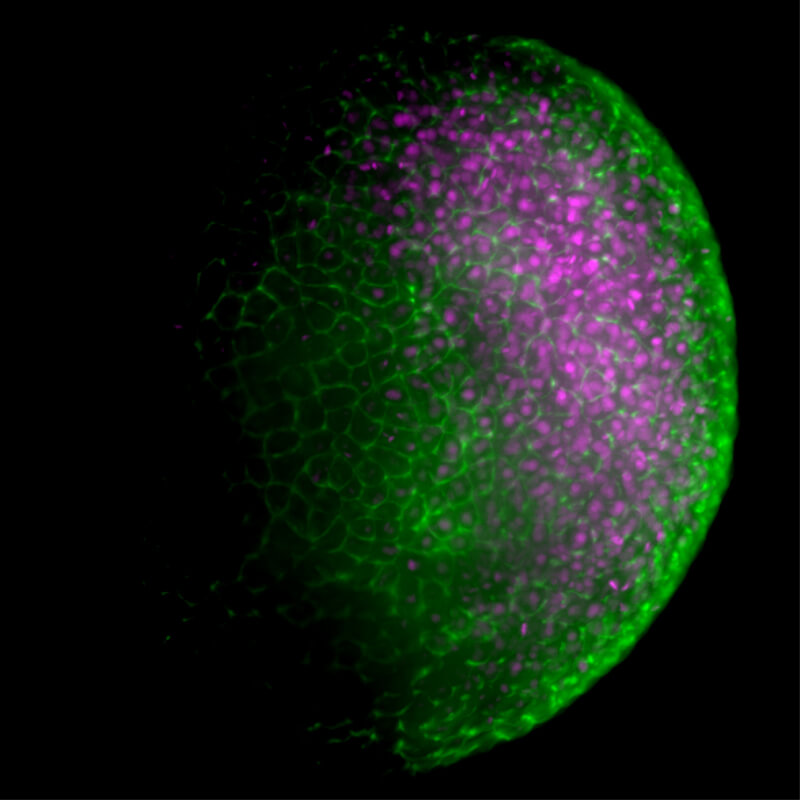
In this early-stage zebrafish embryo, DNA in the nuclei of cells is shown in magenta. The cytoskeleton, in green, shows the structure and boundaries of stem cells. Image courtesy of Gabriel da Silva Pescador/Bazzini Lab.
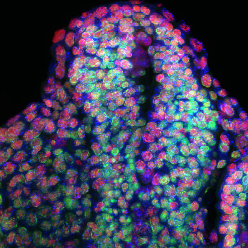
Expression of ribosomal RNA within the neural tube of a mouse embryo. Image courtesy of Karla Terrazas/Trainor Lab.
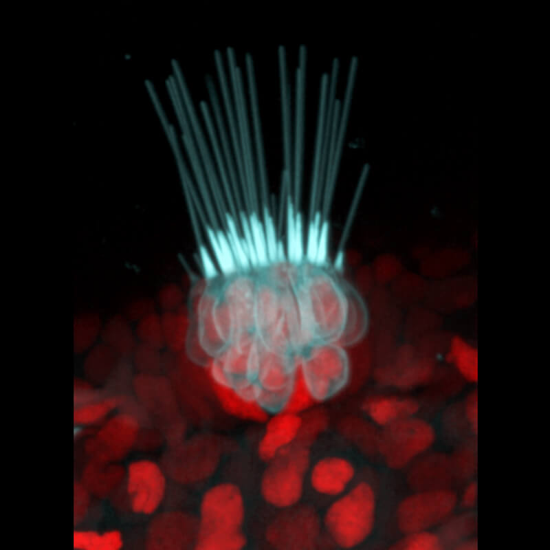
Nature's weathervane - as weathervanes show us the direction of the wind, zebrafish sensory hair cells (cyan) provide them with information on the water movements Image courtesy of Julia Peloggia/Piotrowski Lab.
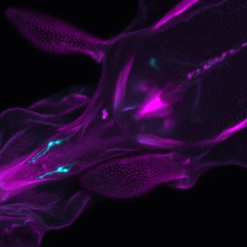
Expression pattern of Drosophila's gustatory receptor-expressing neurons G43a in the proboscis. We labeled the neurons using Gr43aGal4 with UAS-mCD8GFP (cyan in image), no immunostaining. The hairy like structures are the maxillary palps. Image courtesy of Consuelo Perez-Sanchez/Si Lab.
A section through the neural tube of a mouse embryo that shows nascent RNAs, which are the bright spots showing newly made message. Image courtesy of Zainab Afzal/Krumlauf Lab.