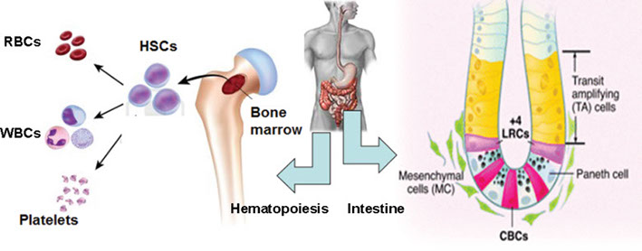
The laboratory of Linheng Li mainly focuses on the hematopoietic and intestinal systems to study stem cell development. The hematopoietic system facilitates functional characterization of stem cells, as bone marrow transplantation experiments can be readily performed. The intestinal system has a well-organized developmental architecture in which stem cell marking and lineage tracing can be used to investigate stem cell behavior. Hematopoietic system research is led by Linheng Li and intestinal system research is led by Senior Research Specialist Xi He.
Stem cells are the key subset of cells in the body that function as ancestor cells producing various types of functionally specialized mature (differentiated) cells in a given tissue, while simultaneously maintaining the capacity to continuously divide and regenerate (self-renewal). This self-renewal process is controlled by intrinsic genetic pathways subject to regulation by extrinsic signals from the microenvironment (or niche) in which stem cells reside. Stem cells play essential roles ranging from embryonic development and organogenesis (fetal stem cells and embryonic stem cells) to tissue homeostasis and regeneration (adult stem cells). Stem cell development is a complex process, and a precise balance is maintained among different cell events including self-renewal, differentiation, apoptosis (cell death), and migration. Loss of this balance tends to lead to uncontrolled cell growth or cell death, thereby developing into various diseases such as cancer or tissue defects.
We mainly focus on the hematopoietic and intestinal systems to study stem cell development. The hematopoietic system facilitates functional characterization of stem cells, as bone marrow transplantation experiments can be readily performed. The intestinal system has a well-organized developmental architecture in which stem cell marking and lineage tracing can be used to investigate how stem cells are maintained by their microenvironment, how stem cells undergo asymmetric/symmetric division to maintain balance between self-renewal and lineage commitment, and what molecular signals are involved in this regulation. To investigate the molecular mechanisms that control stem cell properties, we use the combined approaches described as follows:
1. Molecular basis of multipotentiality of stem cells
What determines the multipotentiality of stem cells is a fundamental question. Analysis of the gene expression profiles during early hematopoietic stem cell development revealed that the step-wise decrease in promiscuity (diversity) for multiple lineage-affiliated genes correlates with a progressive restriction of developmental potential in early hematopoiesis. These results support the hypothesis that stem cells maintain their multipotentiality via a wide-open chromatin structure (Blood 2003).
2. Identification of the hematopoietic stem cell (HSC) niche
Schofield first proposed the hypothesis in 1978 that the niche (the cellular components of the micro environment) plays an essential role in the maintenance of HSCs; however, the HSC niche has remained a mystery since then. By using a Bmpr1a knock-out mouse model, we have identified that spindle-shaped N-cadherin+osteoblastic cells are a key component of the HSC niche. This is the first stem cell niche in the mammalian system to be identified at the cellular level. As an increase in the number of HSCs resulted from an increase in the size of the HSC niche in Bmpr1a mutant mice, BMP signaling controls the HSC number via regulation of the niche size (Nature 2003). The clinical implication of this discovery is the potential to maintain and expand hematopoietic stem cells in vitro.
3. BMP and Wnt signaling yin-yang controls intestinal stem cell (ISC) properties
The molecular mechanism underlying juvenile polyposis syndrome (JPS) caused by defects in Bmpr1a in humans remains largely unknown. Inactivation of Bmpr1a in mice resulted in JPS. We found that the BMP and Wnt signal pathways are antagonistic, ensuring the balanced (yin-yang) control of ISC self-renewal and proliferation versus differentiation. We further propose that BMP signaling inhibits Wnt signaling through either Smad-dependent transcriptional repression or inhibiting β-catenin activity via a PTEN-PI3K/Akt pathway (Nature Genetics 2004).
4. Normal stem cells versus cancer stem cells
In the studies of the Bmpr1a mutant mouse model, we found that the PTEN-controlled PI3K-Akt pathway may mediate a cross talk between BMP and Wnt signaling pathways. We therefore examined the consequences of inactivation of PTEN (a tumor suppressor) in both hematopoietic and intestinal systems. Loss of PTEN leads to enhanced proliferation and mobilization of stem cells, which in turn results in acute myeloid/lymphoid leukemia and intestinal polyposis, respectively. Mechanistic studies of the PTEN deficient mouse model revealed that PTEN plays a critical role in maintaining normal HSCs and preventing leukemia development (Nature 2006); and loss of PTEN results in conversion of normal stem cells into cancer initiating/stem cells. We documented the process of how cancer stem cells initiate tumorigenesis in intestinal polyposis (Nature Genetics 2007).
5. Co-existence of two subpopulations of reserved and primed hematopoietic stem cells (HSCs)
To sustain blood production over a lifetime, primitive HSCs must support daily regeneration of lost cells and must also avoid becoming depleted or exhausted. We have shown how HSCs balance distinct and sometimes opposing needs. We found that the primitive HSC pool contains two sub populations distinguished by the expression level of N-cadherin, an adhesion molecule thought to mediate HSC-niche interaction. Low levels of N-cadherin identify HSCs in a “primed” state ready to support active blood regeneration. Cells with intermediate levels of N-cadherin form a larger pool of “reserved” HSCs adapted to a maintenance role (Cell Stem Cell, 2008). This concept of co-existing quiescent (reserved) and active (primed) stem cells in the same tissue is now well documented in several mammalian tissues including bone marrow (Willson et al., 2008), intestine (Li and Clevers, Science 2010), hair follicle (Fuchs, Cell 2009), and neural system (Pastana et al., PNAS;Mira et al., Cell Stem Cell, 2010).
6. Development of real-time imaging technology for tracing stem cells in vivo or ex vivo
Studying adult stem cell behavior has been limited by a static view of stem cells and their niche structures, due primarily to immunostaining techniques. To overcome this problem, we have developed a new method that allows real-time imaging of stem cell behavior in vivo or ex vivo (Xie et al.,Nature 2009).
7. Expansion of hematopoietic stem cells (HSCs) using a pharmacological approach
Adult stem cells are ready for differentiation into the corresponding tissue cells, which have been exemplified by HSC and bone marrow transplantation. However, HSCs thus far still cannot be expanded robustly in vitro. Based on our genetic study of signaling pathways that regulate HSC self-renewal, we have developed a novel method that allows functional expansion of mouse HSCs.
8. Non-canonical Wnt signaling maintains quiescent reserve HSCs
Previously, Canonical Wnt signaling was the major focus for its role in regulating adult stem cells. We found that Non-canonical Wnt signaling, mediated by Frizzled8 and Flamingo, plays a critical role in maintaining quiescent reserve HSCs in the N-cadherin+endostea niche. A balance between non-canonical and canonical Wnt signaling is critical in maintaining or activating and further proliferating HSCs (Cell 2012).
9. Imprinting mechanism balances HSC quiescence and activation
While external signals are important in regulating HSC properties, how these external signals talk to internal epigenetic and transcription program remains still largely unknown. We found that a unique epigenetic regulation, via imprinting mechanism, at the H19-Igf2 locus, controls the balance of HSCs between quiescence and activation. This work not only brings Igf2-Igfr signaling into play, but also reveals that H19 as a non-coding RNA produces miRNA 475, which in turn suppresses translation of Igf1r, thus preventing quiescent HSC from receiving IGF2 signals, a novel epigenetic regulation mechanism in maintaining a quiescent state of HSCs (Nature 2013).
10. Combinatorial markers isolate ISCs and enhance 3-D organoid culture
Previously, using a reporter mouse model to conduct lineage tracing or sort target stem cell population was the major way to identify stem cells in intestine. Similar to hematopoietic stem cells that can be prospectively isolated using combinatorial markers, we have identified combinatorial surface markers to isolate intestinal and colonic stem cells and substantially improved the in vitro 3-D culture method, thus enabling the formation of organ-like structures from single sorted intestinal and colonic stem cells (Gastroenterology 2013).
Future directions
We are extending our findings of the HSC niche to further dissect the niche signals and investigate how these signals coordinate to regulate stem cell self-renewal. We also are attempting to systematically study the distinct and complementary functions of different types of niche components in the regulation of HSCs. We will map out the different microenvironments required for different stages of hematopoietic lineage commitment and maturation.
As cancer is derived from cancer stem cells, a key to completely curing cancer may depend on whether we can successfully target cancer stem cells (not just the bulk of the tumor mass). To this end, identification and characterization of cancer stem cells will be the essential step. We are engaged in identifying cancer stem cells in hematopoietic and intestinal tissues, followed by characterization of cancer stem cells at cellular and molecular levels. The biggest challenge in treating cancer is drug resistance. Our concept of co-existing quiescent and active stem cells may apply to cancers, in which active cancer stem cells support rapid growth of cancer while quiescent cancer stem cells maintain the seed of malignancy, and thus enrich drug-resistant cells. We propose that to efficiently treat cancer, a new method should focus on targeting both the active and quiescent cancer stem cells.
